
Figure 3.
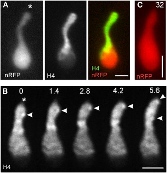
Motility of chromosomes within the nucleus. (A) Chromosomes concentrate at the tip of the nuclear extension, while nRFP is still trapped within the nucleus, indicating that chromosome migration occurs prior to breakage of the envelope. Asterisk indicates leading tip of the nucleus. nRFP: nRFP; H4: histone4-GFP. Bar: 2 μm. (B) Histone4-GFP stains patches within the nucleus that most likely correspond to chromosomes. These patches rapidly migrate towards the tip of the extension (asterisk) at ∼0.25 μm/s (arrowhead). Elapsed time is given in seconds. Bar: 2 μm. (C) nRFP is still present in the nucleus, shown in panel B, indicating that chromosome motility occurred within the closed nucleus. Compare elapsed time given in seconds with series shown in panel B. Bar: 2 μm. Supplementary movie for panel B is given on the EMBO website.