
Figure 6.
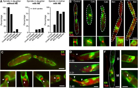
Nuclear migration and NE removal. (A) Quantitative analysis of the effect of nuclear position on NEBD. In control cells, spindles were located in the bud and were never surrounded by an envelope (‘spindle in daughter, without NE'). Treatment with Calcofluor, an inhibitor of chitin synthesis (Herth, 1980), impaired growth and resulted in the formation of spindles within the mother cell. All of the misplaced spindles were surrounded by an apparently intact NE (‘spindle in mother, with NE'). Disruption of MTs by benomyl (MTs inhibited) led to misplaced nuclei that almost always were surrounded by an envelope. Washout of benomyl (MTs released) initiated spindle formation within the closed and misplaced nuclei. No open mitosis was found in dynein mutants. Note that partially opened NEs were counted as ‘with NE'. For further details, see Table I. (B) At standard growth conditions, metaphase spindles were found in the daughter cell and the NE was removed (‘Control'). Treatment with Calcofluor, impaired growth and resulted in misplaced mitotic nuclei with an intact NE (‘Calc'). However, nuclei that had reached the bud did remove the NE. Treatment with the anti-MT drug benomyl (20 μM) impaired nuclear migration and led to condensed nuclei with closed envelopes (‘Ben'). In the absence of MTs, even correctly positioned nuclei were enclosed (‘Ben'). MTs are stained with CFP-Tub1, and NEs are marked with ER-YFP. Bar: 3 μm. (C) Washout of benomyl resulted in the immediate formation of spindles within the closed nuclei. Mitotic nuclei located at the border between mother and daughter cells, as well as in the daughter cell, often showed a partial opening of the NE (arrows indicate gaps in the envelope). Bars: 3 μm in overview and 1 μm in details. (D) After release from benomyl, nuclei that are positioned in the mother cell do not open their envelope. However, the elongating spindle finally disrupted the envelope while it was migrating towards the neck (indicated by asterisk). Note that the condensed chromosomes were still surrounded by small envelopes (arrowheads). Elapsed time is given in minutes:seconds. Spindles are labelled by CFP-Tub1, and envelope are labelled by ER-YFP. Bar: 2 μm. (F) In the absence of dynein, nuclear migration is impaired and nuclei remained in the mother cell (indicated by ‘M', daughter cell is indicated by ‘D'). An apparently intact NE surrounded these misplaced nuclei. Many, partially opened mitotic nuclei were found (see Table I). Red: Spindles labelled by CFP-Tub1, NE marked with ER-YFP. Bar: 2 μm.