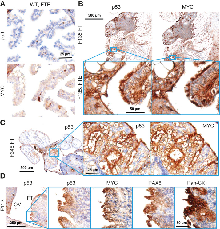
Figure 3.
Molecular histology of the fallopian fimbriae. A, A WT mouse at 12 months of age showing little p53 or MYC staining in the FTE. B, OvTrpMyc fallopian tube at 15 months of age, F135, exhibiting p53 and MYC staining within the FTE. C, An OvTrpMyc mouse at 12 months of age, with vacuolated fallopian fimbriae–positive for p53 and MYC. D, An OvTrpMyc mouse at 15 months of age, with features resembling STIC.

