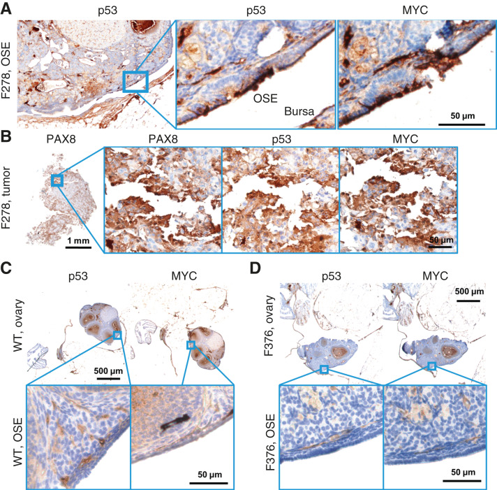
Figure 4.
Molecular histology of the OSE. A, An OvTrpMyc mouse at 16 months of age with clear OSE staining of p53 and MYC. B, The intra-abdominal tumor extracted from the mouse shown in A exhibited PAX8, p53, and MYC staining. C, A WT mouse at 12 months of age is negative for p53 or MYC staining in OSE. D, Example of an OvTrpMyc mouse with a region of negative OSE staining of p53 and MYC, at 12 months of age.

