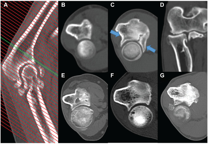
Figure 2.
Radiologic features of proximal radioulnar joint (PRUJ) osteoarthritis on computed tomography images. (A) Level of the cut used to obtain a middle axial view of the PRUJ. (B) PRUJ without osteoarthritic change. (C) Radial notch osteophytes (blue arrows). (D) Radial head osteophyte in coronal view. (E) Joint space narrowing. (F) Subchondral cyst of the radial head. (G) Presence of a loose body.

