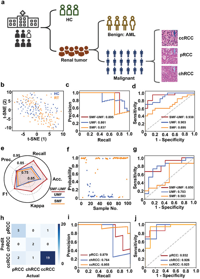
Figure 3.

Three‐step diagnostic models. a) Flowchart of the discrimination model for RCC screening. b–d) Screening of Tumor group: b) t‐SNE visualization of the cluster results of the HC (blue) and tumor (orange) groups; c) AUC of the proposed algorithm on the HC and Tumor groups of precision‐recall (PR) curve; and d) receiver operating characteristic (ROC) curve based on three types of biofluid features. e–g) Identification of tumor type: e) Model evaluation of three types of biofluid features based on the final optimized model, including Recall, Accuracy (Acc.), Kappa, F1, and Precision (Prec.); f) a sample‐probability plot for malignant (orange dots) and benign (blue dots); and g) ROC curve for classifying malignant from benign. h–j) Classification of RCC types: h) confusion matrix for the classification results of the proposed algorithm on the validation dataset (7 pRCC, 3 chRCC, and 20 ccRCC); i) PR curve for three subtypes of RCCs (One vs Rest) i,j) ROC curve of three subtypes of RCCs (One vs Rest).
