
Figure 2.
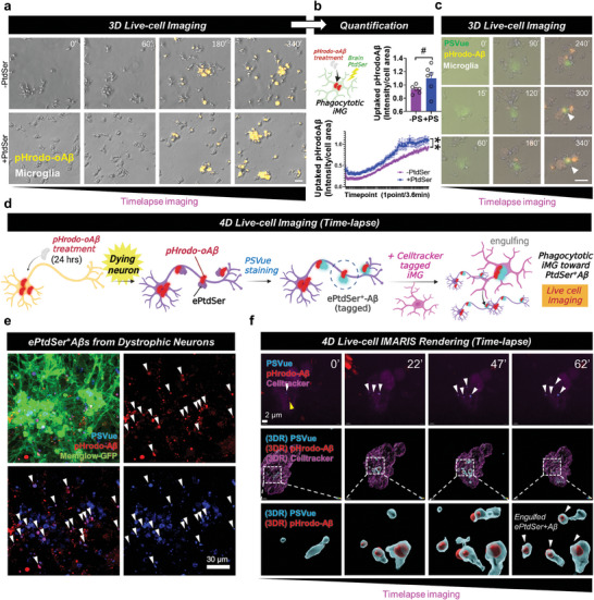
PtdSer improves microglial uptake of Aβ. a,b) Timelapse imaging of microglial uptake of pHrodo‐Aβs with/without PtdSer. Yellow, pHrodo‐Aβ. N = 6 slides for each group; average n = 27.68 cells were tracked per one field of view (scale bar = 20 µm). **p < 0.01, comparison of area under curves; # p < 0.1, independent t‐test. c) PtdSer initializes microglial uptake of pHrodo‐Aβs. Yellow, uptaken pHrodo‐Aβ; green, PSVue. Scale bar = 20 µm. d) Concept of 4D live‐cell imaging to visualize the uptake of ePtdSer+ Aβs by microglia. e) Generation of ePtdSer+ Aβs by dystrophic neurons. Green, Memglow‐GFP, neurons; blue, PSVue; red, pHrodo‐Aβ. f) 4D time‐lapse imaging with 3D rendering using IMARIS software. The internalized pHrodo‐Aβs were colocalized with ePtdSer (ePtdSer+ Aβs). White arrows, internalized ePtdSer+ Aβs; white box, 3D rendering position; blue, PSVue; red, pHrodo‐Aβs; pink, cell‐tracker 647, microglia.