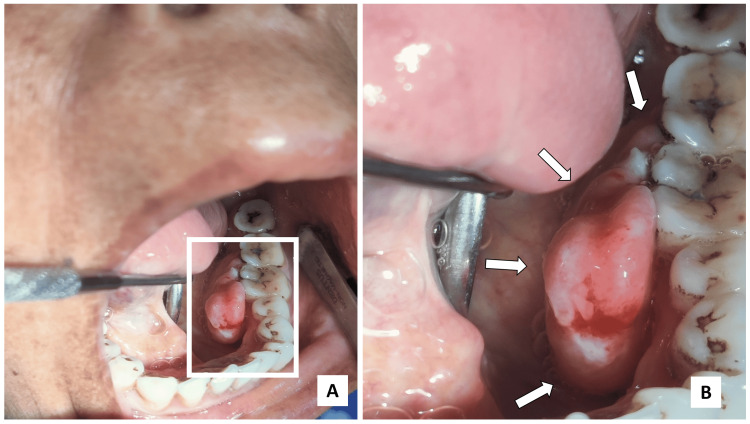
Figure 1. Intraoral examination.
(A) Lobulated, pedunculated mass over the lingual aspect of gingival region w.r.t. 34,35,36,37; (B) Anteroposterior extent of the lesion was from the distal aspect of 34 to mesial aspect of 37 while superoinferiorly it extended from the occlusal surface of regional teeth to depth of the lingual sulcus.
White arrows depict the extent of the growth.

