
Figure 2.
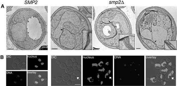
Deletion of SMP2 induces nuclear membrane proliferation and nuclear expansion. (A) Thin section electron microscopy of wild-type (SMP2) or smp2Δ cells grown at 30°C and stained with potassium permanganate. Detail panels show enlargements of areas of the nuclear envelope in smp2Δ cells (highlighted by arrows) that display membrane proliferation. Bars, 0.5 μm. (B) DNA staining of wild-type (SMP2) or smp2Δ knockout cells expressing an intranuclear GFP-reporter (GFP-Pus1) used to depict nuclear structure (‘nucleus'). Cells were grown in selective medium at 30°C, fixed for 30 min and inspected by confocal microscopy. The white arrow points to a dividing yeast cell. Bars, 5 μm.