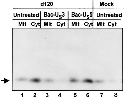
FIG. 6.
Immunoblot showing cytochrome c distribution in HEp-2 cells. HEp-2 cells were either mock infected or infected with the viruses, as indicated, and harvested 18 h after infection. The fractionation procedure was as described in Materials and Methods. Mit, mitochondrial fraction; Cyt, cytosolic fraction.

