
Figure 3.
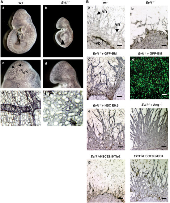
Defects in vascular remodeling and network formation in E9.5 Evi1−/− embryos. (A) Whole-mount PECAM-1-stained wild-type (a) and Evi1−/− (b) embryos at E9.5. Panels (c) and (d) are high-power views of panels (a) and (b), respectively. The Evi1−/− embryo shows pericardial effusion (arrowhead in (b)). The arrows and arrowheads in (c) indicate the remodeled and organized arteries (arrows) and venous vessels (arrowheads). Highly branched small capillaries and network forming vessels were observed in wild type. Evi1−/− counterparts in (d) show no remodeled and organized arteries or veins, and a smaller caliber change in the vessels. (e, f) Yolk sac vascularization. Highly branched small capillaries were evident in wild type (e), while no remodeled or caliber changed vessel was observed in the yolk sac of Evi1−/− (f). (B) Requirement of HSC development for angiogenesis in vitro. P-Sp explants from Evi1−/− (a) and wild-type (b) embryos were dissected at E9.5 and cultured on OP9 cells. P-Sp cultures were stained with anti-PECAM-1 mAbs. The number of hematopoietic cells (round cells) in Evi1−/− P-Sp cultures (Evi1−/−) (b) was significantly less than that observed in wild-type (WT) cultures (a). Defects in the vascular network (vn) in vitro are evident in Evi1−/− P-Sp cultures (b) in comparison to WT cultures (arrows in (a)). The bar indicates 0.1 mm. (c) Development of vascular network stained with anti-PECAM-1 mAbs in Evi1−/− P-Sp culture with HSCs enriched from GFP-positive adult bone marrow (Evi1−/−+GFP-BM). (d) Detection of GFP-positive cells by fluorescence microscopy in (c). The development of vascular network in Evi1−/− P-Sp culture with HSCs from E9.5 WT embryos (Evi1−/−+HSC E9.5) (e) or with the addition of 200 ng/ml recombinant Ang-1 (Evi1−/−+Ang-1). (f) Recombinant Tie2-Fc fusion protein was added with HSCs from E9.5 WT embryos and inhibited the endothelial cell growth of Evi1−/− cells by HSC (Evi1−/−+HSC E9.5/Tie2-Fc). (g) Recombinant CD4-Fc fusion protein was added with HSC and did not inhibit the effect by HSC as a control HSC (Evi1−/−+HSC E9.5/CD4-Fc) (h). The bar indicates 0.1 mm.