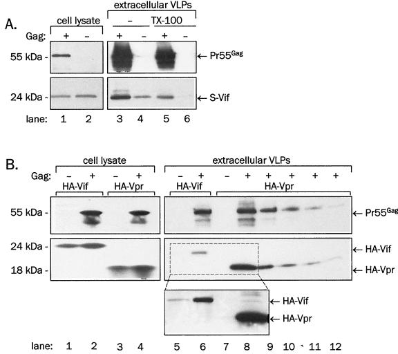
FIG. 5.
Association of Vif or Vpr with Gag VLPs. Lysates of cells coexpressing Gag with HA-Vif or HA-Vpr or lysates of supernatant sediments were resolved on SDS-PAGE. (A) S-tagged Vif was coexpressed with the wild-type Gag in COS-1 cells. An aliquot of sedimented VLPs was treated with 1% Triton X-100 for 15 min at room temperature and resedimented. S-Vif was detected in immunoblotting with anti-Vif rabbit antiserum (bottom), and Gag was detected by reprobing of stripped membranes with anti-CA monoclonal antibody (top). (B) HA-Vif or HA-Vpr were coexpressed with wild-type Gag from the pCITE vector in 293T cells. Lanes 8 to 12: decreasing fractions of supernatant pellet lysate were loaded in the order 1/2, 1/5, 1/10, 1/20, and 1/50. HA-Vif and HA-Vpr were detected in immunoblotting with anti-HA monoclonal antibody (bottom and middle panels), and Gag was detected by reprobing of stripped membranes with anti-CA monoclonal antibody (top). Inset: longer exposure of lanes 5 to 8 indicates background levels of sedimentable Vif and Vpr.

