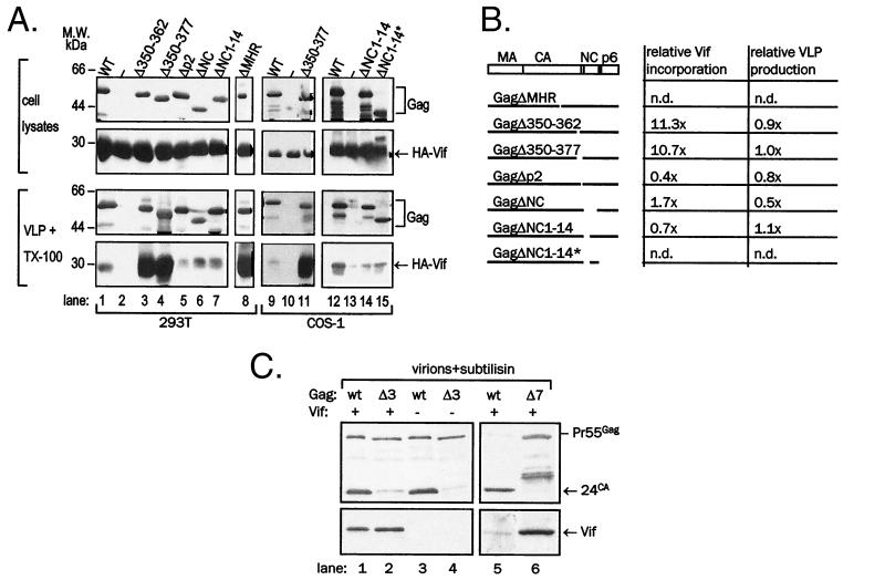
FIG. 7.
Association of Vif with mutant Gag VLPs and virions. (A) Gag was coexpressed with HA-Vif in 293T (lanes 1 to 8) or COS-1 (lanes 9 to 15) cells. VLPs sedimented from supernatants by ultracentrifugation were treated with 1% Triton X-100 to remove background Vif signal, resedimented, and resolved in SDS-PAGE together with a fraction of cell lysates. Vif was detected in immunoblotting with anti-HA monoclonal antibody (second and fourth panels from the top) and Gag was detected by reprobing of stripped membranes with anti-CA monoclonal antibody (top panel and third panel from the top). Gag ΔMHR in lane 8 was detected with AIDS patient sera. Differences in signal intensity among individual panels do not reflect differences in expression levels but rather the variability in film exposure. (B) Schematic representation of Gag deletion mutants used in the study and relative content of Vif in VLPs, normalized to Gag content. (C) Wild-type NL4-3 (wt) and GagΔ350-377 NL4-3 (Δ7) were produced in 293T cells upon transfection with proviral plasmid DNA and treated with subtilisin. Vif was detected in immunoblotting with anti-Vif rabbit antiserum (bottom panel), and Gag was detected by reprobing of stripped membranes with anti-CA monoclonal antibody (top).

