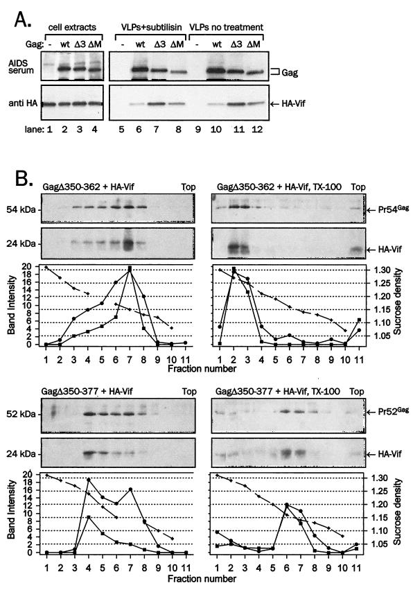
FIG. 8.
Association of Vif with mutant Gag VLP purified by protease treatment or through a sucrose gradient. (A) HA-Vif was coexpressed with wild-type Gag (wt), GagΔ350-362 (Δ3), or GagΔMHR (ΔM) in 293T cells. VLPs collected from supernatants were treated with subtilisin and resolved together with cell lysates on SDS-PAGE. (B) Gag was coexpressed with HA-Vif in 293T cells, and VLPs pelleted from supernatant were treated with Triton X-100 (right panels) or left untreated and resedimented through a 20 to 60% density sucrose gradient overnight. Ten fractions of gradient, starting from the bottom (fraction number 1), as well as the sediment from the bottom of centrifugation tubes representing material denser than 60% sucrose (fractions 11), were collected, resedimented, and analyzed for Vif content in immunoblotting with anti-HA monoclonal antibody (bottom panels) and for Gag by reprobing of stripped membranes with anti-CA monoclonal antibody (top panels). The sucrose density of individual fractions obtained by refractometry or the intensity of Vif and Gag bands obtained by densitometry are plotted below each panel. The solid line with square symbols represents Vif signal intensity; the solid line with circle symbols represents Gag signal intensity; and the dashed line with crossed symbols represents sucrose density.

