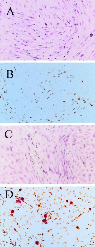
FIG. 2.
The LANA1 protein is expressed in all spindle cell-Converted KSHV-infected DMVEC. (A) High-power phase-contrast photomicrographs of converted spindle cells in a secondary JSC1-infected DMVEC culture at 7 days after 1:10 dilution with fresh uninfected DMVEC. (Crystal violet stain; 40× objective.) (B) Expression of the LANA1 latency protein (brown nuclei) in all spindle cells within two distinct 30- to 80-cell colonies that had formed in a primary infected DMVEC culture at 17 days after addition of JSC1 supernatant virus. LANA1 was detected by IHC with rat anti-LANA1 MAb and peroxidase-DAB chromogen (40× objective.) (C) High-power phase-contrast photomicrograph of a partially spindle cell-converted second-passage DMVEC culture at 24 days after addition of filtered supernatant from a fresh JSC1-infected DMVEC spindle cell culture showing a latently infected spindle cell colony containing a small patch or plaque of rounded cells with typical herpesvirus CPE and some remaining uninfected cuboidal cells. (Crystal violet stain; 40× objective.) (D) Double-label IHC detection of both latent and late lytic cycle KSHV-encoded proteins in a completely spindle cell-converted JSC1-infected DMVEC culture at 3 days after reseeding in a slide chamber dish. Brown chromogen, LANA1 nuclear staining in all latently infected spindle cells (rat anti-LANA1 MAb; peroxidase and DAB). Red/pink chromogen, late lytic cytoplasmic gpK8.1 membrane protein in the subfraction of cells showing rounding and CPE (mouse MAb; phosphatase and Vector red; 40× objective.)

