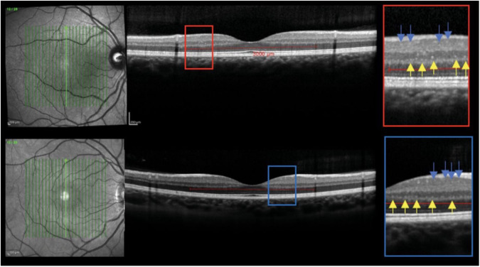
Fig. 3. Optical coherence tomography (OCT) and hyperreflective foci in a patient with relapsing-remitting multiple sclerosis (top OCT) and healthy control (bottom OCT).
The foci in the inner nuclear layer are indicated by yellow arrows, and the foci in the ganglion cell and inner plexiform layer are indicated by the blue arrows. Pengo et al. [19]. observed an association of hyperreflective foci, an indicator of activated and proliferating retinal microglial, on OCT in relapsing-remitting multiple sclerosis compared to healthy controls. Reprinted with permission from Pengo et al. [19]. Retinal Hyperreflecting Foci Associated With Cortical Pathology in Multiple Sclerosis. Neurol Neuroimmunol Neuroinflamm. 2022 May 23;9(4):e1180 under Creative Commons Attribution-NonCommercial-NoDerivatives 4.0 International (CC BY-NC-ND 4.0) License (https://creativecommons.org/licenses/by-nc-nd/4.0/legalcode).

