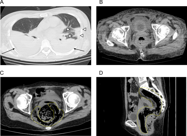
Fig. 1.
A, B CT imaging findings during COVID19 pneumonia. A Bilateral pleural effusion and a consolidation surrounded by ground-glass opacity in the left lung predominance were observed. Arrows indicate the pleural effusion. White arrowheads indicate the consolidation. B Rectum was slightly edematous. C, D CT imaging findings before emergency surgery. C (axial section) and D (sagittal section): free air was observed in the pelvic cavity. A yellow dotted line indicates the free air

