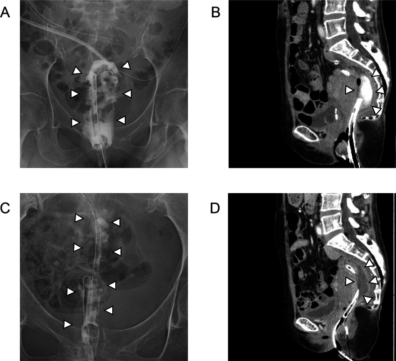
Fig. 3.
A, B Radiographic contrast examination (A) and CT imaging (B) findings after the first surgery. A Transanal drainage tube was added to the pelvic abscess cavity. The abscess cavity was contrasted by gastrografin. B CT image findings indicated a pelvic abscess cavity. C, D Radiographic contrast examination (C) and CT imaging (D) findings before reoperation. C, D Pelvic abscess cavity decreased but persistently remained by radiographic contrast examination (C) and CT (D). White arrowheads indicate the abscess cavity

