
Figure 4.
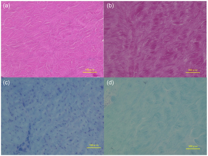
Histological evaluation of the meniscus. (a) Hematoxylin and eosin staining ×200; histologic observations in the intact MM group showing normal cell density, normal proteoglycan staining and regular alignment of collagen fibre fascicles. (b) Hematoxylin and eosin staining ×200; histologic observations in the UM group showing irregular alignment of collagen fibre fascicles. (c) Toluidine blue staining ×200; histologic observations in the intact MM group showing normal proteoglycan content. (d) Toluidine blue staining ×200; histological observations in the UM group showing a reduction in cell density and proteoglycan content. UM, untreated medial meniscal ramp lesions.