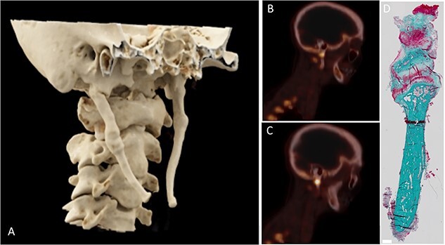
Figure 1.

3D reconstruction from CT image, sagittal [18F]NaF PET/CT images, and microscopic detail image of patient 2. The styloid process was elongated bilaterally (A) with the right styloid process causing symptoms. Increased [18F]NaF uptake was observed at the base of the styloid process (B, C) with a pseudoarthrosis being visible in the histological detail of the styloid process (D).
