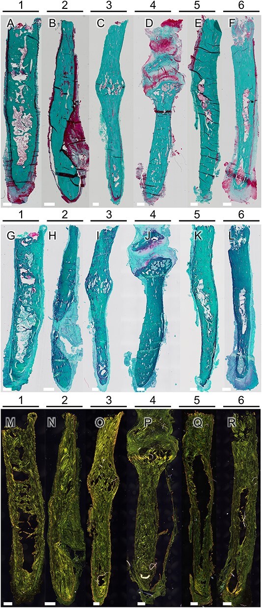
Figure 2.

Microscopic images of full biopsies generated for each sample after Masson-Goldner staining (A–F). Bars for scale are 500 μm. No epiphysis is visible, which indicates no abnormal site of bone growth. In addition, Safranin-O staining revealed remnants of cartilaginous tissue in one sample (G), while the other samples (H-L) showed no evidence of cartilage within the bone tissue, except for one sample (J) exhibiting cartilage formed at a pseudarthrosis, possibly following a fracture of the styloid process. One sample exhibited reddish staining (L) in the surrounding soft tissue, which may be a sign of proteoglycans or inflammation. Picrosirius red staining (M–R) was utilized to enhance the circularly polarized light (CPL) signal for imaging collagen alignment. In all samples, a plywood-like structure, including osteonal structures, was observed.
