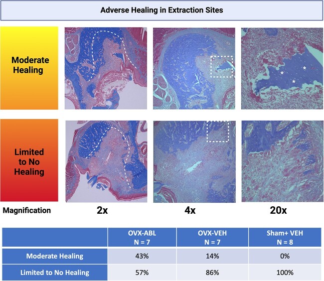
Figure 5.
Histopathological spectrum of osteonecrosis in extraction sites. Histology of extraction sockets with moderate, limited, or no healing at 2×, 4×, and 20× magnification. In the 2× view, the dashed lines indicate the socket borders. In the 2× view of the sample with limited to no healing, the socket is filled entirely with connective tissue without signs of inflammatory infiltrates. The dashed, square boxes in the 4× view indicate the region of magnification in the accompanying 20× view. The arrows in the 4× view of the sample with limited healing indicate bone apposition characterized by thickening or sclerosis of the basal aspect of the maxilla. In the 20× views of the samples with moderate and limited healing, asterisks mark the isolated sequestra of necrotic bone filled with numerous empty osteocytic lacunae. A chi-squared test revealed no significant difference in the number of samples with moderate or limited to no healing between treatment groups. (p = .1151).

