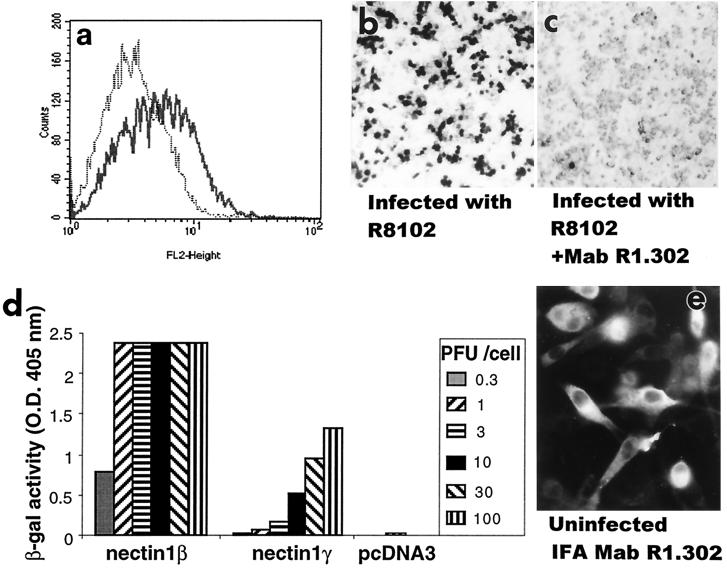
FIG. 4.
Nectin1γ mediates HSV infectivity. A clonal transformant of J1.1-2 cells expressing nectin1γ was stained with MAb R1.302 (a) and infected with R8102 virus (b and c). (c) Prior to infection, cells were exposed to MAb R1.302 directed against nectin1 (1:25-diluted ascitic fluid) or with an irrelevant control antibody to human herpesvirus 6 (1:25-diluted ascitic fluid or purified IgGs [0.16 μg/μl] [not shown]) for 2 h at 4°C. Infection was monitored as β-Gal activity, by staining with 5-bromo-4-chloro-3-indolyl-β-d-galactopyranoside (X-Gal). (d) Stable cell lines expressing nectin1β, nectin1γ, or pcDNA3 were infected with R8102, at increasing multiplicities of infection ranging from 0.3 to 100 PFU/cell. Infection was monitored as β-Gal activity. O.D. 405 nm, optical density at 405 nm. (e) Intracellular distribution of nectin1γ detected by indirect immunofluorescence assay (IFA) with MAb R1.302. J1.1-2 cells expressing nectin1γ were fixed with 4% paraformaldehyde for 15 min and permeabilized with 0.1% Triton X-100 for 10 min.

