Table 1.
Parameters of the test dataset.
| Modality | Parameter | MPR of Scan Range | |
|---|---|---|---|
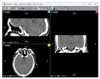
| Data 1 | CT | Slice Spatial Resolution: 512 × 512 Slice Quantity: 121 Pixel Spacing: 0.4336 mm\0.4336 mm Thickness: 1.0 mm ROI from radiological description: An oval-shaped nodular shadow can be seen on the inner side of the left optic nerve behind the orbital ball, suspected to be a hemangioma |
|
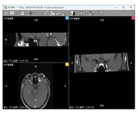
| Data 2 | MRI | Slice Spatial Resolution: 288 × 384 Slice Quantity: 18 Pixel Spacing: 0.625 mm\0.625 mm Thickness: 3.0 mm ROI from radiological description: An oval-shaped nodular shadow can be seen on the inner side of the left optic nerve behind the orbital ball, suspected to be a hemangioma |
|
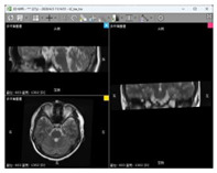
| Data 3 | MRI | Slice Spatial Resolution: 512 × 512 Slice Quantity: 15 Pixel Spacing: 0.4296875 mm\0.4296875 mm Thickness: 3.5 mm ROI from radiological description: Nodular shadow on the left side of the saddle area, considering the possibility of pituitary adenoma |
|
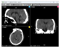
| Data 4 | CT | Slice Spatial Resolution: 512 × 512 Slice Quantity: 25 Pixel Spacing: 0.430 mm\0.430 mm Thickness: 5 mm ROI from radiological description: postoperative cerebellar changes, abnormal structural disturbances in the cerebellar region, occipital bone showing postoperative changes |
|
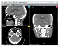
| Data 5 | CT | Slice Spatial Resolution: 512 × 512 Slice Quantity: 177 Pixel Spacing: 0.401 mm\0.401 mm Thickness: 1 mm ROI from radiological description: cerebral softening foci in the left part of the brainstem, demyelinating changes in the cerebral white matter |
|
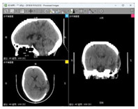
| Data 6 | CT | Slice Spatial Resolution: 512 × 512 Slice Quantity: 30 Pixel Spacing: 0.46289 mm\0.46289 mm Thickness: 5 mm ROI from radiological description: large area of bone defect in the left temporoparietal bone adjacent to the left temporalis muscle, edema in the left temporoparietal lobe of the brain, formation of softening lesions, and slight swelling of the temporalis muscle. |
|
