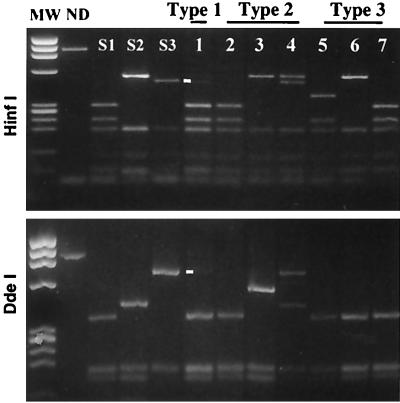
FIG. 2.
RFLP analysis of vaccine-derived PV strains. The 3′-terminal parts of the genomes of strains isolated from vaccinees were amplified by RT-PCR and digested with restriction enzymes HinfI and DdeI (RFLP-3D-3′ assay). The restriction profiles of the reference Sabin 1, 2 and 3 (S1, S2, and S3) strains and of some type 1 (lane 1), type 2 (lanes 2 to 4), and type 3 (lanes 5 to 7) strains isolated from vaccinees and identified by serotyping are shown. The profiles shown in lanes 1 to 7 correspond to recombinant genomes. Whereas most profiles correspond to a single genotype, lanes 1 and 4 show mixtures of two different genotypes. Lanes 3 (HinfI) and 5 (DdeI) show hybrid restriction fragments indicating the locations of recombinant sites in the amplified fragment. Faint minor bands (lane 1) corresponding to S3 genomic fragments are indicated by dashes. Other minor bands resulting from incomplete digestion were observed in a few lanes (lanes 1 and 2). MW, HaeIII-digested fragments of bacteriophage φX174 DNA serving as molecular weight markers; ND, not digested.

