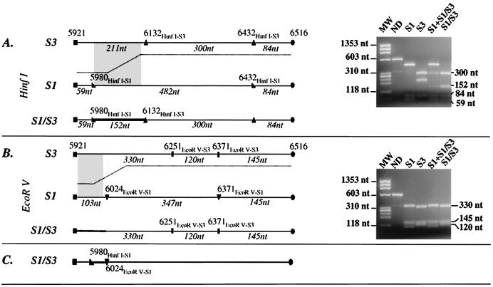
FIG. 6.
RFLP pattern (RFLP-3D3) and restriction map of the amplicon carrying the recombination site of an S1/S3 recombinant genome. Restriction profiles obtained from the reference Sabin 1 and 3 strains (S1 and S3), from the original mixed isolate (S1+S1/S3), and from a plaque-purified recombinant virus (S1/S3) are shown. Nucleotide positions of the amplicon extremities and of the restriction sites on the PV genome are indicated according to Sabin 1 strain numbering. Genomic regions believed to include the recombination junction are indicated by grey zones and bold lines. Lengths of restriction fragments (in nucleotides [nt]) are indicated. Molecular weight markers (MW) are φX174 DNA HaeIII digested fragments. ND, nondigested. (A) HinfI restriction map and profiles on an agarose gel; (B) EcoRV maps and profiles; (C) localization of the recombination junction of the S1/S3 genome inferred from profiles in panels A and B.

