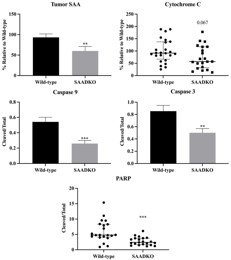
Figure 4.
SAADKO tumors showed lower SAA levels, have reduced apoptosis signaling, and increased DNA repair. Caspase 3 and caspase 9 were analyzed with an uncorrected unpaired t-test. Tumor SAA was analyzed with an unpaired t-test with Welch’s correction. Cytochrome c and PARP were analyzed with a Mann–Witney test. Tumor SAA, and Caspase 3/9 data show the mean ± SEM, while cytochrome c and PARP data show the median with IQR (n = 7). ** p < 0.01 WT vs. SAADKO, *** p < 0.001 WT vs. SAADKO. Representative Western blot images can be found in Supplementary File S4 (Figure S13).

