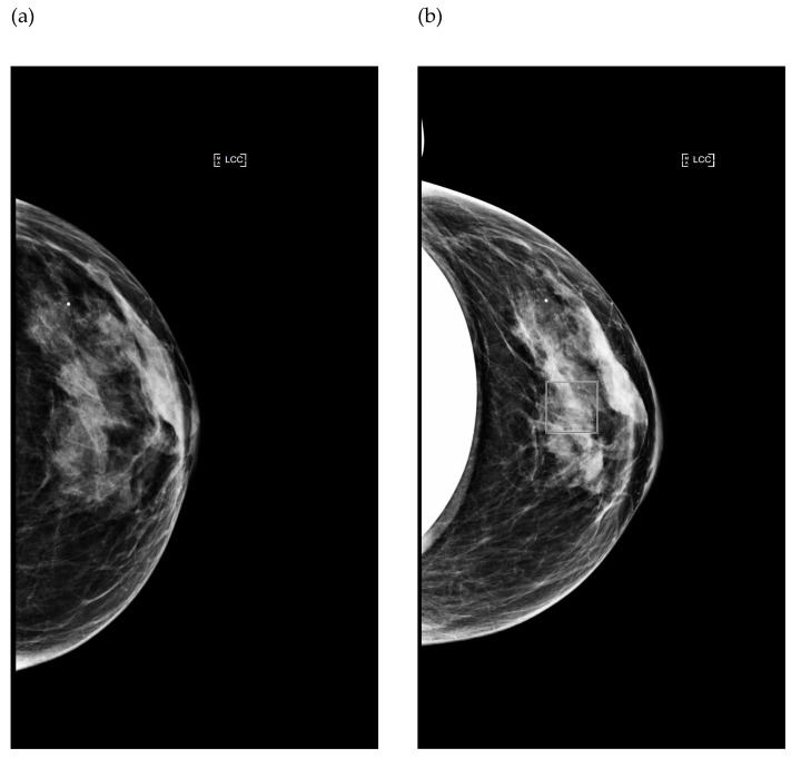
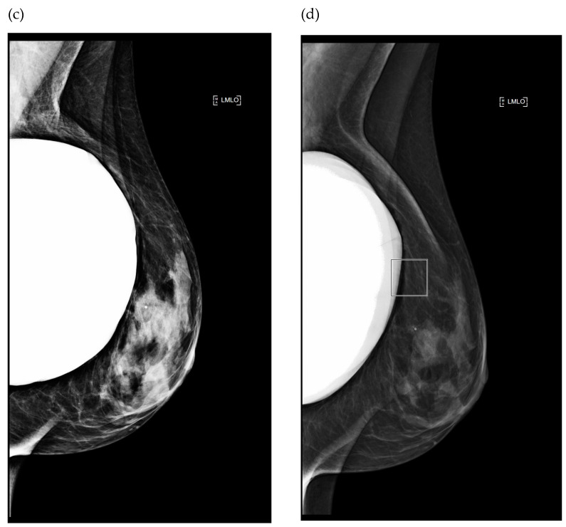
Figure 4.
Image comparison between two different mammograms performed in the same patient during follow-up, using the manual (a,c) and fully automatic (b,d) modes. (a,b) Left cranio caudal projection; (c,d) Left medio-lateral oblique projection. The squares displayed on images indicate the location of the AEC sensor for the automatic mode.


