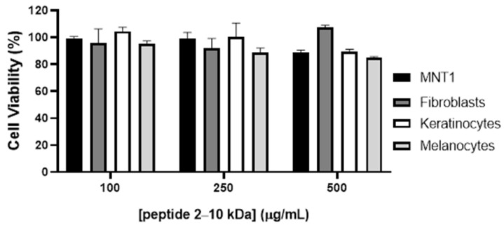
Figure 3.
Cytotoxicity of the peptides (2–10 kDa) in melanoma cell line MNT1 and normal dermal fibroblasts, keratinocytes, and melanocytes. Cells were exposed to increasing concentrations of the peptide for 48 h and viability was evaluated with the MTS assay. Bars represent the average ± standard deviation.

