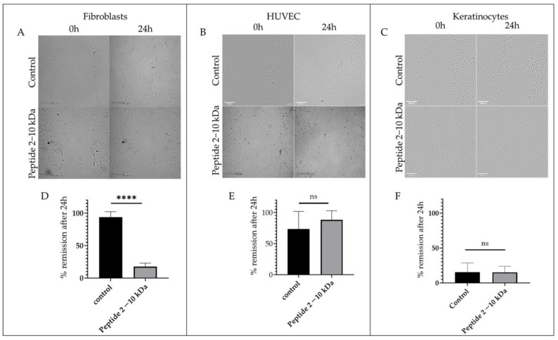
Figure 6.
Wound healing assay. Representative images of the wound healing assay at 0 h and 24 h after incubation with 1% (v/v) ethanol (control) or 250 µg/mL of peptides (2–10 kDa) in (A) normal dermal fibroblasts, (B) HUVEC, and (C) keratinocytes. Scale bars correspond to 200 µm. Percentage of wound scratch closure (% remission) after 24 h incubation with 1% (v/v) ethanol (control) or 250 µg/mL of peptides (2–10 kDa) in (D) normal dermal fibroblasts, (E) HUVEC and (F) keratinocytes. Bars represent the mean ± SEM of at least two independent experiments. **** p value < 0.0001, ns—statistically not significant.

