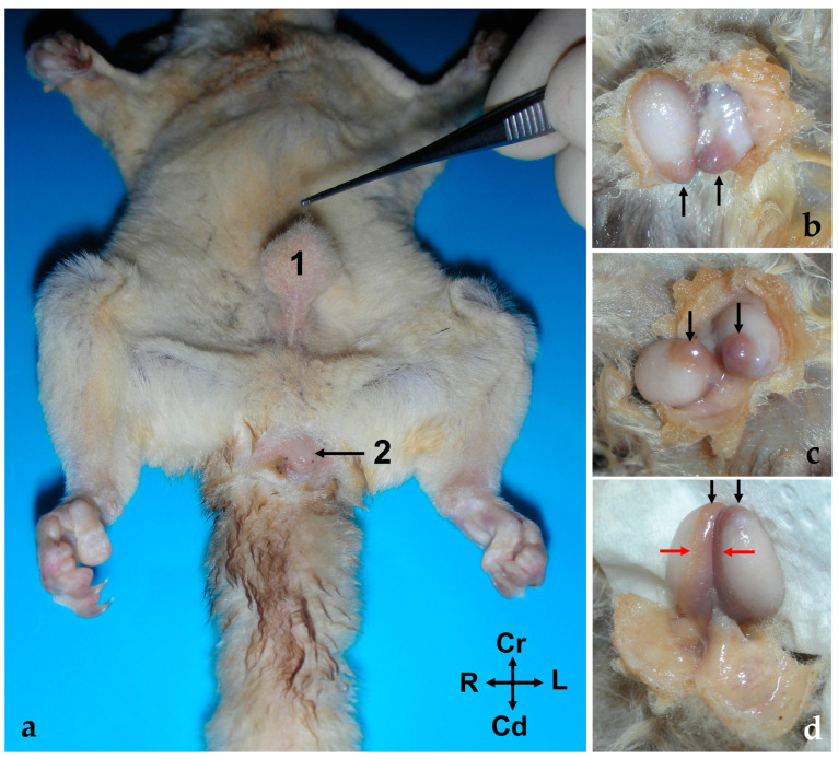
Figure 1.
(a) Ventral view of the abdominal wall of a male sugar glider (fresh specimen); 1, the scrotum; 2, the cloacal orifice. (b–d) Images providing a detailed view of the testes and epididymis in their natural position. The scrotum has been incised for better visibility. The black arrows indicate the tail of the epididymis, while the red arrows point to the body of the epididymis. (b) Cranial view, (c) ventral view, and (d) caudal view. All photographs maintain the same orientation as indicated in image (a).

