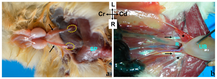
Figure 3.
(a) Ventral view of a male sugar glider’s abdominal wall. The extra-abdominal course of the deferent ducts is indicated by black arrows, leading towards the superficial inguinal ring, represented by a discontinuous oval shape; Te, testes (b) ventral view of a male sugar glider’s abdominal cavity, with the digestive viscera displaced cranially. The intra-abdominal trajectory of the deferent ducts, again indicated by black arrows, leads towards the dorsal and cranial prostate. The urinary bladder (UB) has been reflected in a caudal direction to reveal its dorsal surface and the beginning of the prostate gland. The right deferent duct is demarcated by a red dotted line, while the terminal segment of the right ureter is indicated by a blue dotted line. Both males were fresh specimens. R, Rectum; SP, symphysis pubica; Te, testes; UB, urinary bladder; ✱, lateral vesical ligaments with round ligaments of the bladder. The orientation is the same in both images.

