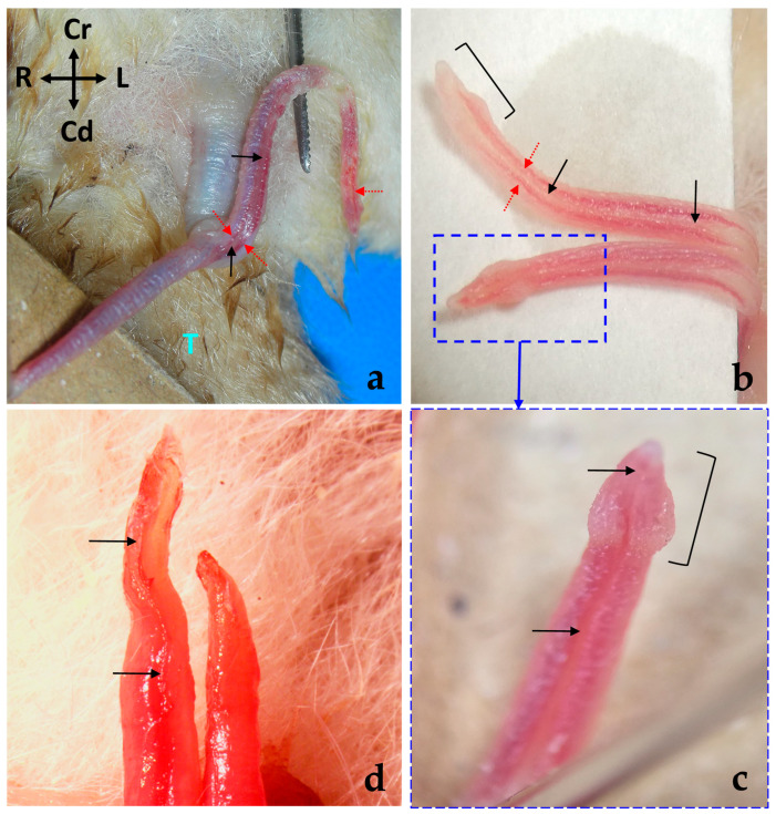
Figure 6.
(a) Ventral view of the externalized penis of a male sugar glider in situ, exteriorized through the cloacal orifice. Fresh specimen. The left half of the free part has been displaced to observe its medial wall. (b) Medial wall of the bifurcated end of the penis, showing venous sinuses (red arrows). (c) Inset of (b) displaying the cone-shaped termination of the tip ( ] ) of the bifurcated penis, with a prominent widening at its base. Note the urethral groove (black arrows) continues up to the tip. (d) Tips of the bifurcated penis from a castrated male, showing the absence of the widened conical termination.

