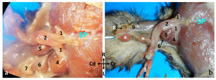
Figure 8.
Examination of a fresh specimen. (a) Ventral view of a male sugar glider’s penis in situ. The anatomical structures on its left side have been dissected. (b) Ventral left view of a male sugar glider’s penis. The prepuce has been incised and the penis has been extracted from the preputial sac. 1, Crus penis; 2, corpus penis; 3, pars libera penis; 4, left ischiocavernosus muscle; 5, left bulbospongiosus muscle; 6, left bulbourethral gland I; 7, left bulbourethral gland II; 8, cloacal cavity; 9, rectum; ✱, paracloacal glands. SP, Symphysis pubica; both images have the same orientation.

