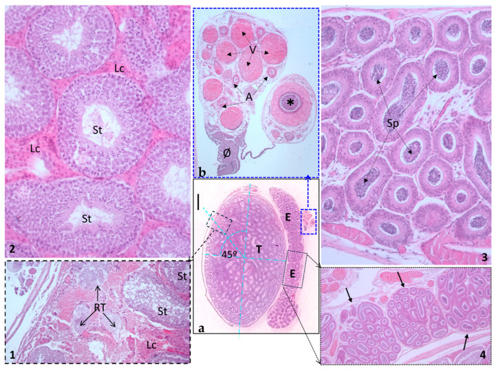
Figure 10.
Testicle and epididymis. (a) A low-magnification section showing the testicle (T) and epididymis (E). Scale bar represents 1 mm. (1) An inset of (a) displays the rete testis, positioned at a 45º angle from the perpendicular axes (as depicted in (a)), from which the efferent duct transports spermatozoa to the epididymis. (2) Seminiferous tubules (St) teeming with numerous interstitial or Leydig cells (Lc). Note the distinct associations within the seminiferous tubules, exhibiting various stages of sperm cell development. (3) Histological section of the epididymis, revealing numerous cross-sections of the same epididymal duct filled with spermatozoa (Sp). (4) The black inset displays the characteristic lobuli epididymidis (black arrows) at a low magnification (objective 4×). (b) The blue inset in panel (a) provides a closer look at the spermatic cord and its components: ✱, deferent duct surrounded by a thick circular muscular layer; A, sections of the testicular artery; V, veins of the plexus pampiniformis; Ø, cremaster muscle (distal portion).

