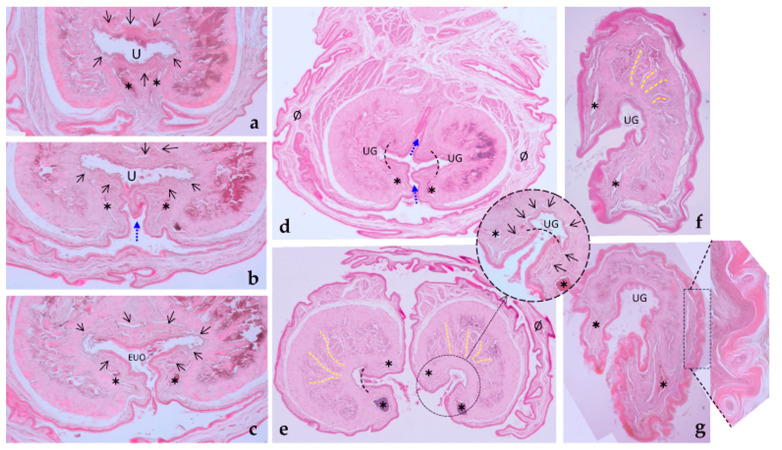
Figure 11.
Penile histology. (a) The penile urethra is surrounded by the corpus spongiosum (black arrows) and the corpus cavernosum. (b) A ventral groove (blue arrow) deepens and almost reaches the penile urethra, just before the external urethral orifice. (c) The ventral groove reaches the urethra, which opens to the exterior, forming the external urethral orifice (EUO). (d) The groove continues dorsally (blue arrows) to form the bifurcated tip of the penis, and the continuation of the urethra creates two urethral grooves (UG) in a medial position of each part. Note the presence of two venous sinuses (✱) flanking the groove walls. The penis is protected by the mucosa of the prepuce (Ø). (e) The bifurcated penis is now separated from the prepuce, and each half is independent. The inset shows the urethral groove (UG), with urothelium at the groove’s bottom (the limits are indicated by the dotted curved line) and cutaneous mucosa forming the groove walls. This entire structure is surrounded by the corpus spongiosum (black arrows) and two venous sinuses on either side of the groove (one dorsal and one ventral (✱)). In the corpus cavernosum, yellow lines indicate dense connective tissue septa with a radial arrangement between blood-filled spaces. (f) The termination of the bifurcated penile tip, at the beginning of the conical formation. The corpus cavernosum is clearly visible, suggesting the absence of a glans. (g) An oblique section of the penile termination reveals cornified spines on the surface (inset). In both (f,g), the corpus cavernosum is evident, further supporting the absence of a glans.

