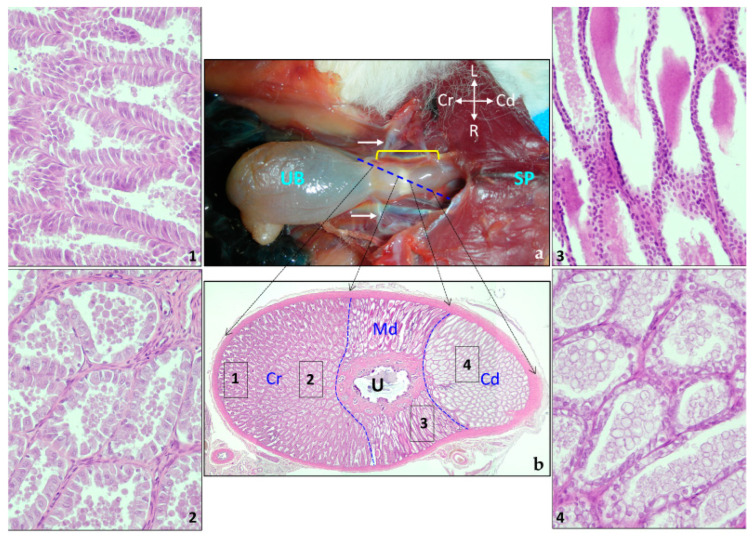
Figure 12.
Prostate gland. (a) Macroscopic ventral view of the prostate gland in situ. The three parts of the prostate (yellow bracket) can be distinguished. White arrows, deferent ducts; SP, symphysis pubica; UB, urinary bladder. (b) Oblique histologic section of the prostate at the level of the blue dotted line in (a). The central image is a composition of three images made with the 4× objective and the numbers represent different areas of the cranial portion (Cr), middle (Md), and caudal part (Cd) easily distinguishable by their morphology. Photographs 1–4 represent the glandular tissue from the areas indicated in (b) to display their glandular epithelium and the secretion appearance (made with the 40× objective). U, Urethra.

