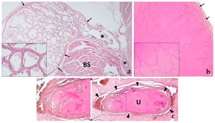
Figure 13.
Histology of the bulbourethral glands. (a) Image showing bulbourethral gland I at low magnification to observe its tubulo-alveolar morphology and its secretion duct (✱). The entire gland is externally lined by striated muscle (black arrows) arranged in two perpendicular layers. The inset shows at higher magnification (40× objective) the morphology of the glandular epithelium made up of a single layer of cubic cells. BS, bulbospongiosus muscle. (b) Bulbourethral gland II, also of tubulo-alveolar nature, filled with acidophilic secretion. Two layers of striated muscle also appear surrounding the gland capsule. The inset shows at higher magnification (40× objective) the appearance of low cubic secretory cells arranged forming a single layer of cells lining the alveoli. The secretion, strongly acidophilic, presents some granules of more intense staining. (c) Section of the penile urethra showing the point of discharge of the ducts of the bulbourethral glands pouring their secretion into the lumen of the urethra. The inset shows in greater detail the discharge of the left duct. Note the presence of the spongy body surrounding the urethra (black arrowheads).

