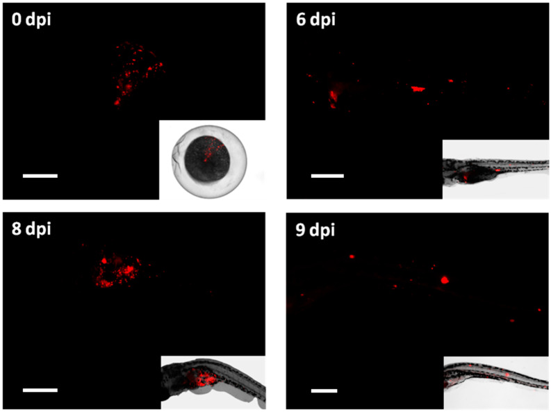
Figure 3.
Fluorescent microscopy imaging of Mycobacterium tuberculosis infection in zebrafish larvae. Images taken with a fluorescent stereomicroscope of 0, 6, 8 and 9 dpi M. tuberculosis-DsRed infected larvae are shown together with the bright field image of the same embedded larvae. Image of 0 dpi embryo represents a successful injection of fluorescent bacteria into the yolk. At 6, 8 and 9 dpi bacteria showed dissemination throughout the body of the larvae and the formation of bacterial aggregates. Scale bar: 200 micrometer.

