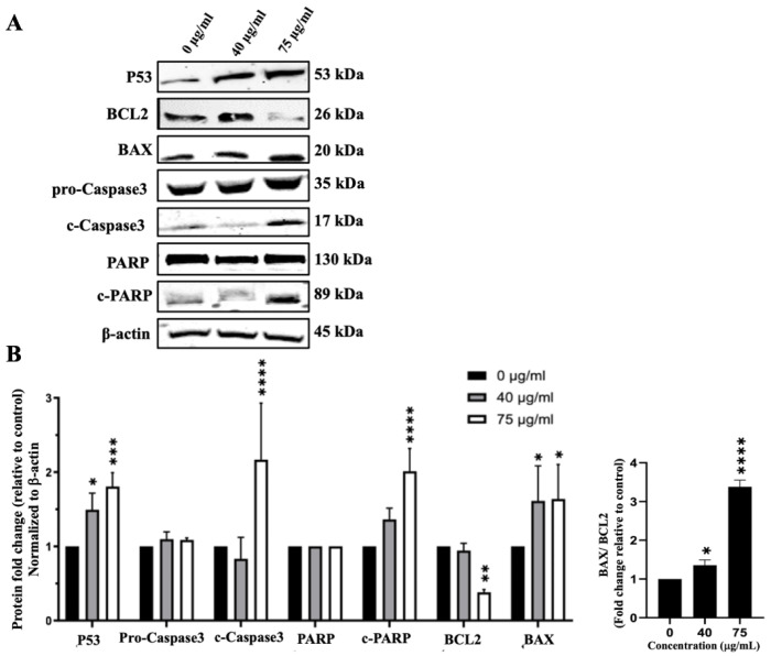
Figure 7.
IVL DCM induces the apoptosis of A549 cells. A549 cells were treated with the indicated concentrations of IVL DCM for 48 h. (A) P53, BCL2, BAX, Caspase 3, c-Caspase 3, PARP, c-PARP protein levels as detected by immunoblotting of A549 cell lysates. β-actin was immunoblotted as a loading control (B). Quantification of the bands in (A). Bar graphs of band intensity of target proteins normalized to the intensity of the loading control β-actin expressed as fold change of the vehicle control and represented as the mean ± SEM of three independent experiments (n = 3). The right panel of (B) shows the ratio of BAX/BCL2 (n = 3). * p < 0.05, ** p < 0.01, *** p < 0.001, and **** p < 0.0001.

