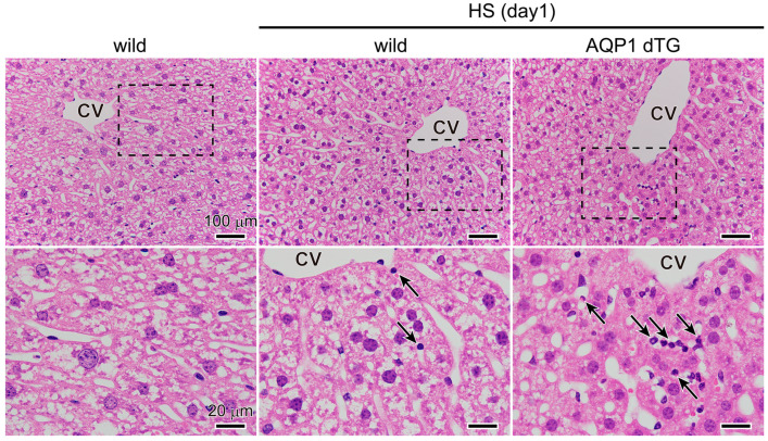
Figure 6.
Morphological comparisons of the liver. Representative images of livers after hematoxylin–eosin staining in non-heat-exposed and heat-exposed wild-type and Tie2-Cre/LNL-AQP1 dTG mice. High-magnification images of each rectangle are shown at the bottom. Leukocytes were observed after heat exposure (indicated by arrows). CV: central vein of hepatic lobules.

