Figure 8.
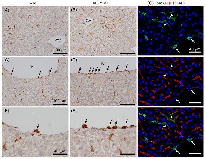
Iba1 immunostaining in the liver. A red-brown Iba1-ir was observed in the sinusoids, suggesting the presence of Kupffer cells, and it did not show a remarkable difference between wild-type (A) and Tie2-Cre/LNL-AQP1 dTG (B) mice. Iba1-ir (black arrows) was also observed in small- and medium-sized vessels, such as interlobular vessels (IVs), and was more prominent in Tie2-Cre/LNL-AQP1 dTG (D,F) mice than in wild-type mice (C,E). (E,F) show high-magnification images of Iba1-ir on IV. (G) Multiple staining for Iba1 (green) and AQP1 (red) was performed to determine whether Iba1+ cells expressed AQP1. Iba1+ cells were observed in Kupffer cells (arrowhead) and vascular adherent monocytes/macrophages (white arrows) but did not co-localize with AQP1-ir. Blue 4′,6-diamidino-2-phenylindole (DAPI) staining indicates nuclei. CV, central vein; IV, interlobular vessel; scale bars, 100 μm (A–D) and 40 μm (E–G).

