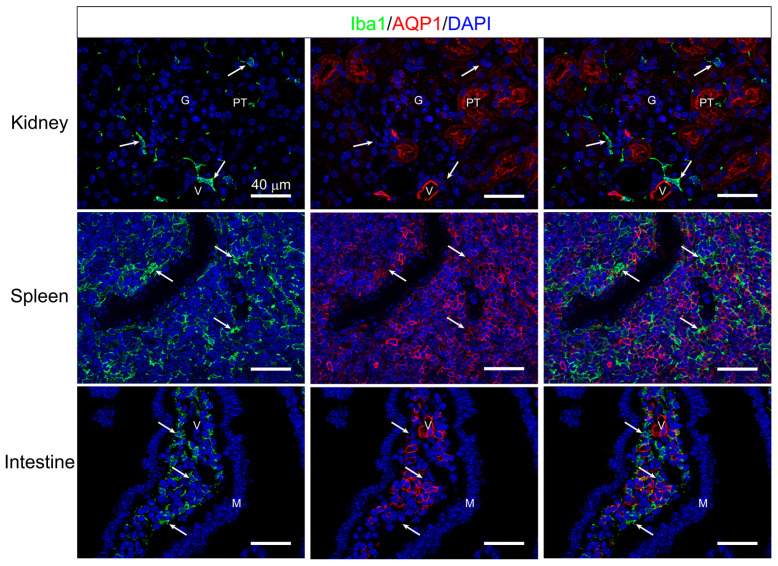
Figure 9.
Multiple immunostaining of Iba1 and AQP1. Multiple staining of pan-monocyte/macrophage markers, Iba1 (green) and AQP1 (red), were performed in the kidney (upper), spleen (middle), and intestine (bottom), in addition to the liver (see Figure 8) to eliminate the expression of AQP1 in monocyte and macrophage lineage cells in Tie2-Cre/LNL-AQP1 dTG mice. The AQP1- ir was observed in the proximal tubules (PTs) and vasculature (V), including the glomeruli (G). However, Iba1-ir (arrow) was localized in the stroma and did not co-localize. In the spleen, the area of red pulp is recognized by many AQP1-ir molecules. While Iba1-ir (arrow) was also recognized in the red pulp, it did not co-localize. The AQP1-ir in the intestine was observed in the lamina propria but not in the mucous epithelia (M). Some were likely stromal cells, whereas others formed vasculature-like (V) structures. Although many Iba1-irs (arrow) were also recognized in the lamina propria, they did not co-localize. Blue DAPI staining indicates nuclei. Scale bar: 40 μm.

