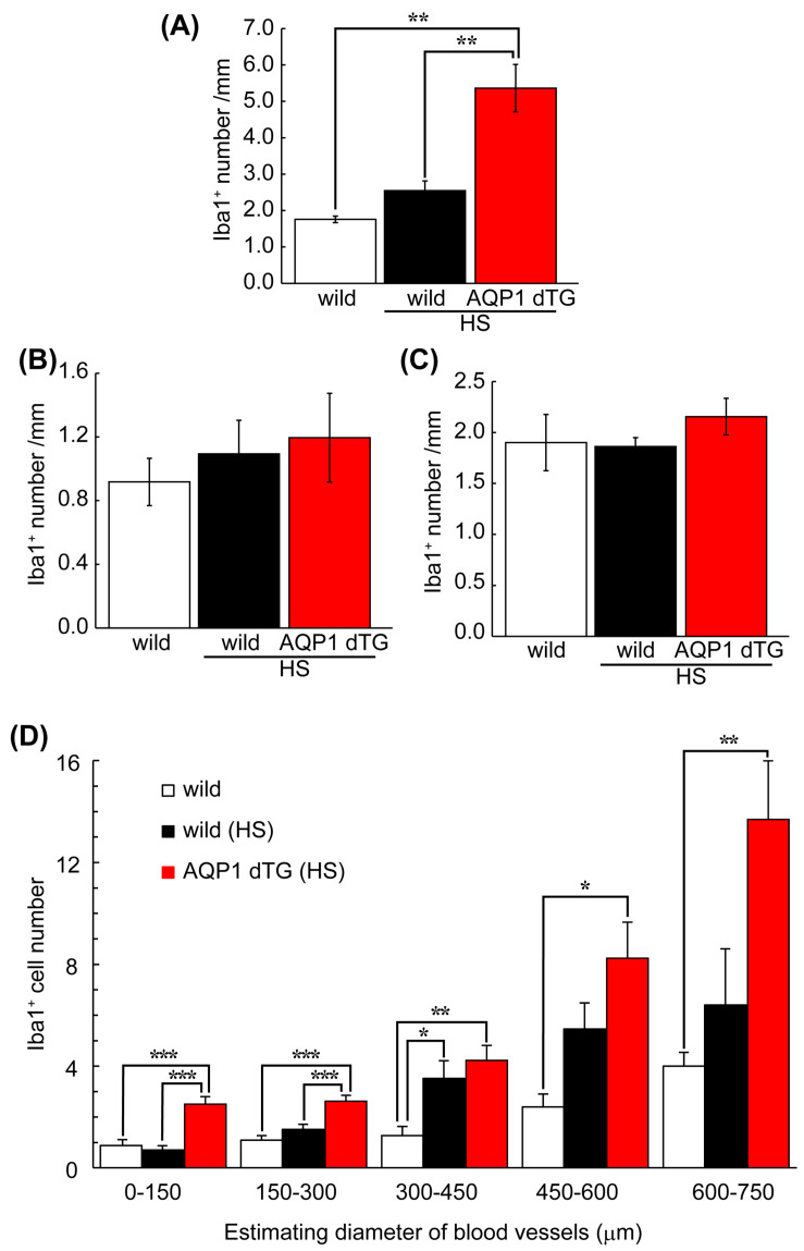
Figure 10.
Comparison of Iba1-positive numbers on the blood vessels of the liver. Vasculature-adherent Iba1-positive cells (Iba1+) were counted in 30 vessels of the liver (A) and brain (B) and 20–24 vessels of the kidney (C) in each animal. The results were expressed as the mean number and length of the vessels (mm). (A) The number of Iba1+ cells was significantly higher in Tie2-Cre/LNL-AQP1 dTG mice (n = 5) than in wild-type mice, both with (n = 4) and without (n = 3) heat exposure. The numbers did not differ between the brain and kidneys. (D) The number of Iba1+ cells in the liver was used to estimate the diameter of each blood vessel, calculated from the circumferential length. While Iba1+ numbers in Tie2-Cre/LNL-AQP1 dTG mice were greater in all vessel sizes, the diameters of vessels measuring 0–150 and 150–300 μm differed significantly from those in wild-type mice after heat exposure. The data are expressed as mean ± SE. * p < 0.05, ** p < 0.01, *** p < 0.001 (Tukey’s post hoc test).

