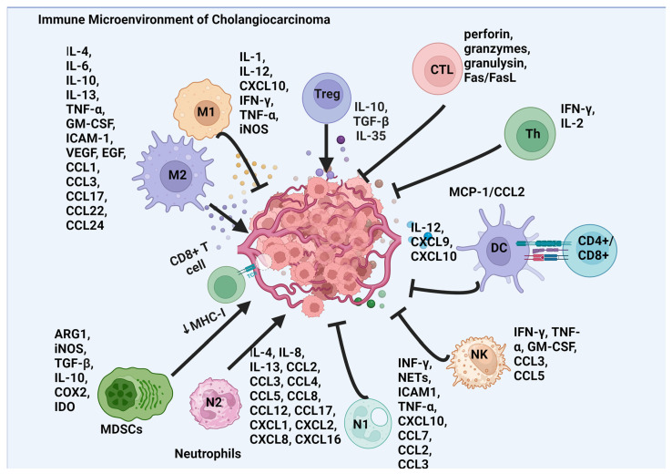
Figure 1.
Schematic of the intricate immune microenvironment in CCA, mapping the interplay between different immune cells and their associated mediators. The diagram displays the tumor in the center, surrounded by various immune cells: M1 and M2 macrophages, which release pro-inflammatory and anti-inflammatory cytokines, respectively; T-regulatory cells (Tregs), which produce immunosuppressive cytokines; cytotoxic T lymphocytes (CTLs), which secrete cytotoxic molecules; helper T cells (Th), which aid in immune response modulation; dendritic cells (DCs), which present antigens; natural killer (NK) cells, which release cytotoxic substances; and myeloid-derived suppressor cells (MDSCs), which contribute to tumor growth and immune evasion. Additionally, the graphic outlines the complex network of cytokines and chemokines, such as interleukins, tumor necrosis factors, and chemotactic cytokines (CCLs and CXCLs), highlighting the multifaceted interactions that contribute to the cancer’s immune environment. Abbreviations: arginase 1 (ARG1), chemokine (C-C motif) ligand (CCL), CXC motif ligand (CXCL), cyclo-oxygenase (COX), epidermal growth factor (EGF), interferon (IFN), indoleamine 2,3-dioxygenase (IDO), intracellular adhesion molecule-1 (ICAM-1), granulocyte–macrophage colony-stimulating factor (GM-CSF), major histocompatibility complex (MHC), neutrophil extracellular traps (NETs), tumor necrosis factor (TNF), transforming growth factor-β (TGF-β), vascular endothelial growth factor (VEGF).

