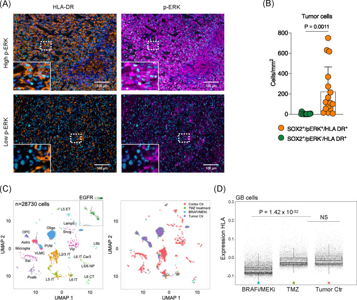
Figure 6. MAPK/ERK pathway modulates antigen presenting process in glioma cells.
(A) Multiplex immunofluorescence analysis of human GB tumor. Representative images of high p-ERK tumor (top) and low p-ERK tumor (bottom). Scale bar = 100 μm. (B) Quantified phenotypic analysis of multiplex immunofluorescence across 16 GB patient samples. Bar graph showing SOX2+HLA-DR+ cells comparing p-ERK− and p-ERK+ cells. (C) Single cell analysis from slice cultivated human GB tumor treated with temozolomide (left) and BRAFi/MEKi (right). (D) Differential expression of human leukocyte antigen (HLA) in slice cultivated human GB sample without treatment or temozolomide and BRAFi/MEKi treated (P = 1.42 × 10−52).

