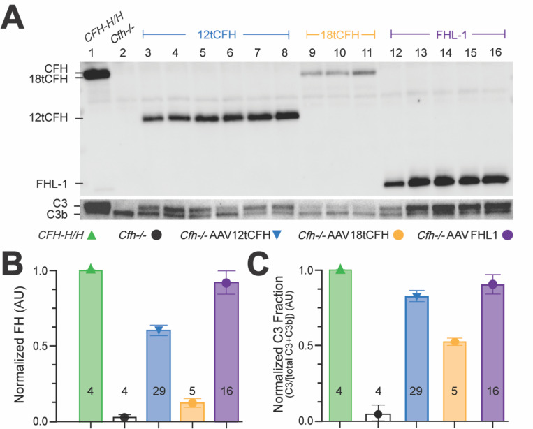
Figure 2. Systemic, AAV-mediated replacement of CFH in Cfh−/− mice drives hepatic tCFH expression and reduces fluid phase C3b through restoration of the APC.
(A) Representative Western blots showing circulating plasma levels of 12tCFH, 18tCFH, and FHL-1 in Cfh−/− mice treated by AAV-mediated CFH replacement. (B) Bar graph indicating normalized CFH expression in each cohort (± AAV treatment). The number of mice analyzed for each cohort is shown at the base of each bar. (C) Bar graph of normalized C3 fraction calculated as the C3 fraction of the total C3 + C3b for each animal and normalized against CFH-H/H, C3G-negative control mice. The number of mice analyzed for each cohort is shown at the base of each bar.

