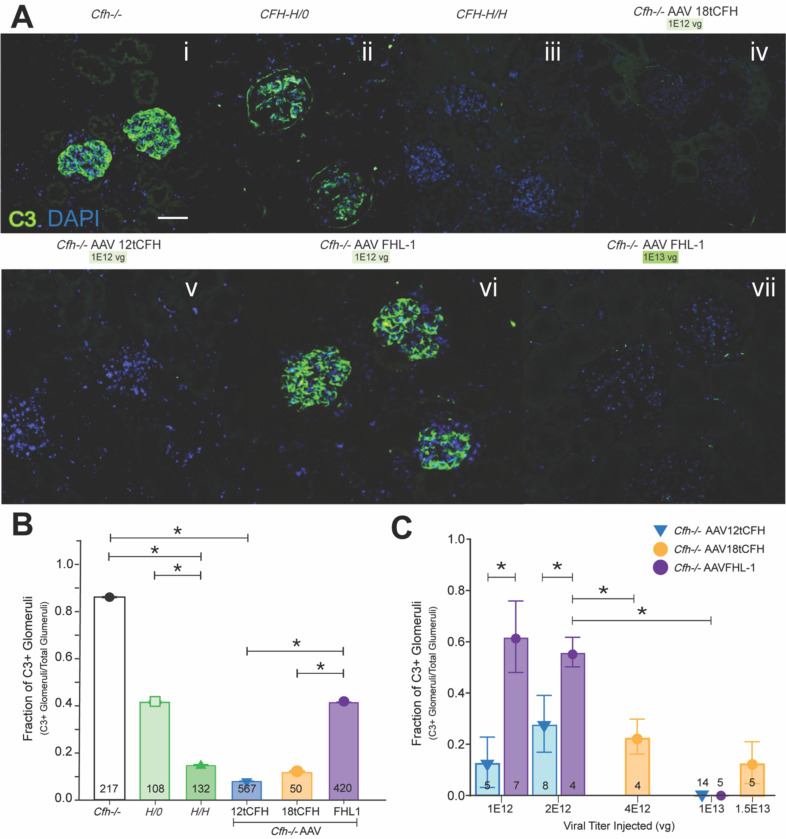
Figure 3. AAV-mediated CFH replacement reduces C3 deposit accumulation and resolves C3G.
(A) Representative immunofluorescent confocal imaging of glomeruli from kidney sections of mice according to genotype indicated and AAV treatment (viral titer delivered) as shown. Scale bar is 100 microns. (B) Bar graph indicating the fraction of C3-positive (C3+) glomeruli out of the total glomeruli for which confocal imaging was obtained. The number of glomeruli analyzed for each cohort is shown at the base of each bar. Cfh−/− n=9; CFH-H/0 n=6; CFH-H/H n=9; Cfh−/− AAV12tCFH n=29; Cfh−/− AAV18tCFH n=9; and Cfh−/−AAVFHL-1 n=16. Unpaired t-test was used for comparison between treatment cohorts, *p<0.01. (C) Bar graph analyzing only AAV-treated Cfh−/− mice and assessing treatment as a function of the fraction of C3+ glomeruli out of the total glomeruli imaged. The number of mice in each treatment cohort is shown at the base of each bar. Unpaired t-test was used for comparison between treatment cohorts, *p<0.01.

