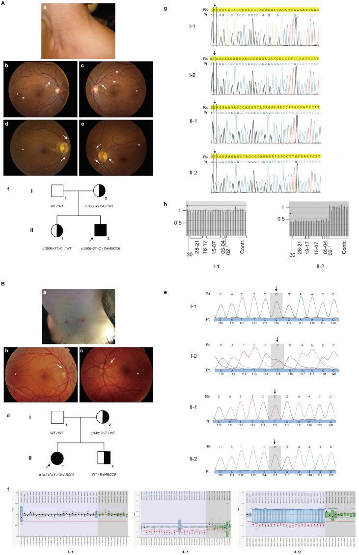
Figure 2.
Clinical characteristics, pedigrees and molecular findings of the proband families. Panel (A): proband 1. (a) Papular skin lesions in the lateral neck at the time of diagnosis; (b–e) white-light funduscopy of the proband (b,c) and his mother (d,e). Peau d’orange (asterisk), comet tails (open arrows) and angioid streaks (closed arrows) are shown. (f) Pedigree of the family of proband 1 (II-2, arrowed). The ABCC6 genotype is indicated for each individual (WT = wild type). (g) Electropherograms of the ABCC6 direct sequencing results for individuals I-1, I-2, II-1 and II-2. The location of the affected nucleotide at position c.3506 + 2 is arrowed. Due to the presence of a heterozygous whole-gene deletion, only a single peak is observed in the proband, in contrast to overlapping peaks in his mother and sister. (h) MLPA results of the proband (II-2) and his father (I-1). Every bar is the ratio is the result of 1 probe pair PCR product. From left to right: ratio for the ABCC1 probe, ABCC6 exon 30, exon 28-21, exon 18-17, exon 15-7, exon 5, exon 4 and exon 2, followed by 12 bars representing the control probes (contr.). The ABCC1 control probe is indicated with an asterisk. All ratios for the control samples are ~1, indicating that there is no deletion or duplication present. In the father (I-1), all ratios also equal to 1 indicate that no deletion is present; in the proband (II-1), all ABCC6 probes and the ABCC1 probe have a ratio of ~0.5, confirming the presence of a heterozygous ABCC6 whole-gene deletion. The deletion of ABCC1 is commonly associated with ABCC6 deletions and confirms that also exon 1 of ABCC6—for which no probe is present in the MLPA kit—is deleted; the ABCC1 deletion however has no phenotypic consequences as was previously described [17]. Panel (B): proband 2. (a) Papular skin lesions in the lateral neck at the time of diagnosis; (b,c) white-light funduscopy of the proband. Peau d’orange (asterisk) and angioid streaks (closed arrows) are shown. (d) Pedigree of the family of proband 2 (II-1, arrowed). The ABCC6 genotype is indicated for each individual (WT = wild type). (e) Electropherograms of the ABCC6 direct sequencing results for individuals I-1, I-2, II-1 and II-2. The location of the affected nucleotide at position c.3421 is arrowed. (f) MLPA results of the proband (II-1), her father (I-1) and her brother (II-2). The ratio results of the probe pair PCR products are indicated by the dots; black dots: the ratio lies within the 95% confidence interval (CI) of the reference sample population, indicating the absence of a deletion or duplication; red: the ratio lies out of the 95% CI and over the arbitrary borders (0.7 to 1.3 by default), indicating the presence of a deletion or duplication. Whiskers: 95% CI for sample value (test or reference). Boxes: 95% CI in reference sample population (by default). Blue: compared to test probes; green: compared to reference probes. From left to right: ratio for the TSC2 probe, ABCC1 probe, ABCC6 exon 30, exon 28-21, exon 18-17, exon 15-7, exon 5, exon 4 and exon 2, followed by 10 bars representing the control probes (reference*). All ratios for the negative control sample are ~1, indicating that there is no deletion or duplication. In the father (I-1) all ratios also equal to 1, indicating that no deletion is present; in the brother (II-2) and the proband (II-1), all ABCC6 probes and the ABCC1 probe again have a ratio of ~0.5, confirming the presence of a heterozygous ABCC6 whole-gene deletion.

