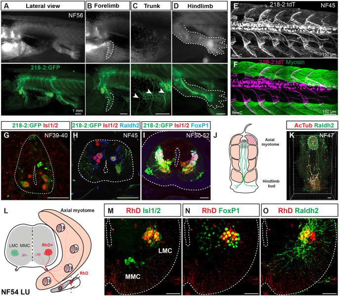
Figure 3. Linking motor neuron molecular profiles to anatomical projection pattern during frog metamorphosis.
A-D. GFP-labeled motor neurons in a limbed NF56 218-2:GFP tadpole (A) innervate the forelimb (B), trunk (C), or hindlimb (D), in line with the expected innervation patterns of the molecular populations detected at this stage. Scale bar, 1 mm.
E-F. Motor neurons in free-swimming NF45 218-2:GFP:tdT tadpoles are distributed throughout the spinal cord and extend axonal arbors into the myotomal cleft, as shown by myosin heavy chain immunohistochemistry (green; F). Scale bar, 150 μm.
G-I. GFP-labeled motor neurons at the limb levels in 218-2:GFP tadpoles at NF39–40 (G), NF45 (H) and NF50–54 (I) express the pan-motor neuron marker Isl1/2 (red). At NF45 and at NF50–52, a Raldh2/FoxP1-positive population (blue) of motor neurons, consistent with their LMC identity, also expresses GFP. Scale bar, 50 μm.
J-K. A pioneering population of LMC motor neurons at free-swimming stage NF47 expresses the LMC marker Raldh2 (green) and their axons, marked with the acetylated-α-Tubulin antibody (red), extend to the developing limb area prior to limb bud emergence. Scale bar, 50 μm.
L. Schematic of medial and lateral motor column innervation patterns and the location of the rhodamine dextran application. Scale bar, 50 μm.
M-O. Retrograde labeling with rhodamine dextran (RhD, red) marks a population of laterally positioned motor neurons that co-express MN (Isl1/2, green; M) and LMC (FoxP1/Raldh2, red; N/O) markers. Scale bar, 50 μm.

